

A cataract is a clouding of the lens in the eye that affects vision. Most cataracts are related to aging. Cataracts are very common in older people. By age 80, more than half of all Americans either have a cataract or have had cataract surgery.
A cataract can occur in either or both eyes. It cannot spread from one eye to the other.

The lens is a clear part of the eye that helps to focus light, or an image, on the retina. The retina is the light-sensitive tissue at the back of the eye.
In a normal eye, light passes through the transparent lens to the retina. Once it reaches the retina, light is changed into nerve signals that are sent to the brain.
The lens must be clear for the retina to receive a sharp image. If the lens is cloudy from a cataract, the image you see will be blurred.
Yes. Although most cataracts are related to aging, there are other types of cataract:

Normal vision

The same scene as viewed by a person with cataract
The lens lies behind the iris and the pupil (see diagram). It works much like a camera lens. It focuses light onto the retina at the back of the eye, where an image is recorded. The lens also adjusts the eye's focus, letting us see things clearly both up close and far away. The lens is made of mostly water and protein. The protein is arranged in a precise way that keeps the lens clear and lets light pass through it.
But as we age, some of the protein may clump together and start to cloud a small area of the lens. This is a cataract. Over time, the cataract may grow larger and cloud more of the lens, making it harder to see.
Researchers suspect that there are several causes of cataract, such as smoking and diabetes. Or, it may be that the protein in the lens just changes from the wear and tear it takes over the years.
Age-related cataracts can affect your vision in two ways:
The term "age-related" is a little misleading. You don't have to be a senior citizen to get this type of cataract. In fact, people can have an age-related cataract in their 40s and 50s. But during middle age, most cataracts are small and do not affect vision. It is after age 60 that most cataracts steal vision.
The risk of cataract increases as you get older. Other risk factors for cataract include:
Wearing sunglasses and a hat with a brim to block ultraviolet sunlight may help to delay cataract. If you smoke, stop. Researchers also believe good nutrition can help reduce the risk of age-related cataract. They recommend eating green leafy vegetables, fruit, and other foods with antioxidants.
If you are age 60 or older, you should have a comprehensive dilated eye exam at least once every two years. In addition to cataract, your eye care professional can check for signs of age-related macular degeneration, glaucoma, and other vision disorders. Early treatment for many eye diseases may save your sight.
The most common symptoms of a cataract are:
Cataract is detected through a comprehensive eye exam that includes:
Your eye care professional also may do other tests to learn more about the structure and health of your eye.
The symptoms of early cataract may be improved with new eyeglasses, brighter lighting, anti-glare sunglasses, or magnifying lenses. If these measures do not help, surgery is the only effective treatment. Surgery involves removing the cloudy lens and replacing it with an artificial lens.
A cataract needs to be removed only when vision loss interferes with your everyday activities, such as driving, reading, or watching TV. You and your eye care professional can make this decision together. Once you understand the benefits and risks of surgery, you can make an informed decision about whether cataract surgery is right for you. In most cases, delaying cataract surgery will not cause long-term damage to your eye or make the surgery more difficult. You do not have to rush into surgery.
Sometimes a cataract should be removed even if it does not cause problems with your vision. For example, a cataract should be removed if it prevents examination or treatment of another eye problem, such as age-related macular degeneration or diabetic retinopathy. If your eye care professional finds a cataract, you may not need cataract surgery for several years. In fact, you might never need cataract surgery. By having your vision tested regularly, you and your eye care professional can discuss if and when you might need treatment.
If you choose surgery, your eye care professional may refer you to a specialist to remove the cataract.
If you have cataracts in both eyes that require surgery, the surgery will be performed on each eye at separate times, usually four to eight weeks apart.
Many people who need cataract surgery also have other eye conditions, such as age-related macular degeneration or glaucoma. If you have other eye conditions in addition to cataract, talk with your doctor. Learn about the risks, benefits, alternatives, and expected results of cataract surgery.
There are two types of cataract surgery. Your doctor can explain the differences and help determine which is better for you:
After the natural lens has been removed, it often is replaced by an artificial lens, called an intraocular lens (IOL). An IOL is a clear, plastic lens that requires no care and becomes a permanent part of your eye. Light is focused clearly by the IOL onto the retina, improving your vision. You will not feel or see the new lens.
Some people cannot have an IOL. They may have another eye disease or have problems during surgery. For these patients, a soft contact lens, or glasses that provide high magnification, may be suggested.
As with any surgery, cataract surgery poses risks, such as infection and bleeding. Before cataract surgery, your doctor may ask you to temporarily stop taking certain medications that increase the risk of bleeding during surgery. After surgery, you must keep your eye clean, wash your hands before touching your eye, and use the prescribed medications to help minimize the risk of infection. Serious infection can result in loss of vision.
Cataract surgery slightly increases your risk of retinal detachment. Other eye disorders, such as high myopia (nearsightedness), can further increase your risk of retinal detachment after cataract surgery. One sign of a retinal detachment is a sudden increase in flashes or floaters. Floaters are little "cobwebs" or specks that seem to float about in your field of vision. If you notice a sudden increase in floaters or flashes, see an eye care professional immediately. A retinal detachment is a medical emergency. If necessary, go to an emergency service or hospital. Your eye must be examined by an eye surgeon as soon as possible. A retinal detachment causes no pain. Early treatment for retinal detachment often can prevent permanent loss of vision. The sooner you get treatment, the more likely you will regain good vision. Even if you are treated promptly, some vision may be lost.
Talk to your eye care professional about these risks. Make sure cataract surgery is right for you.
Cataract removal is one of the most common operations performed in the United States. It also is one of the safest and most effective types of surgery. In about 90 percent of cases, people who have cataract surgery have better vision afterward.
A week or two before surgery, your doctor will do some tests. These tests may include measuring the curve of the cornea and the size and shape of your eye. This information helps your doctor choose the right type of IOL.
You may be asked not to eat or drink anything 12 hours before your surgery.
At the hospital or eye clinic, drops will be put into your eye to dilate the pupil. The area around your eye will be washed and cleansed.
The operation usually lasts less than one hour and is almost painless. Many people choose to stay awake during surgery. Others may need to be put to sleep for a short time.
If you are awake, you will have an anesthetic to numb the nerves in and around your eye.
After the operation, a patch may be placed over your eye. You will rest for a while. Your medical team will watch for any problems, such as bleeding. Most people who have cataract surgery can go home the same day. You will need someone to drive you home.
Itching and mild discomfort are normal after cataract surgery. Some fluid discharge is also common. Your eye may be sensitive to light and touch. If you have discomfort, your doctor can suggest treatment. After one or two days, moderate discomfort should disappear.
For a few days after surgery, your doctor may ask you to use eyedrops to help healing and decrease the risk of infection. Ask your doctor about how to use your eyedrops, how often to use them, and what effects they can have. You will need to wear an eye shield or eyeglasses to help protect your eye. Avoid rubbing or pressing on your eye.
When you are home, try not to bend from the waist to pick up objects on the floor. Do not lift any heavy objects. You can walk, climb stairs, and do light household chores.
In most cases, healing will be complete within eight weeks. Your doctor will schedule exams to check on your progress.
Problems after surgery are rare, but they can occur. These problems can include infection, bleeding, inflammation (pain, redness, swelling), loss of vision, double vision, and high or low eye pressure. With prompt medical attention, these problems can usually be treated successfully.
Sometimes the eye tissue that encloses the IOL becomes cloudy and may blur your vision. This condition is called an after-cataract. An after-cataract can develop months or years after cataract surgery.
An after-cataract is treated with a laser. Your doctor uses a laser to make a tiny hole in the eye tissue behind the lens to let light pass through. This outpatient procedure is called a YAG laser capsulotomy. It is painless and rarely results in increased eye pressure or other eye problems. As a precaution, your doctor may give you eyedrops to lower your eye pressure before or after the procedure.
You can return quickly to many everyday activities, but your vision may be blurry. The healing eye needs time to adjust so that it can focus properly with the other eye, especially if the other eye has a cataract. Ask your doctor when you can resume driving.
If you received an IOL, you may notice that colors are very bright. The IOL is clear, unlike your natural lens that may have had a yellowish/brownish tint. Within a few months after receiving an IOL, you will become used to improved color vision. Also, when your eye heals, you may need new glasses or contact lenses.
If you have lost some sight from cataract or cataract surgery, ask your eye care professional about low vision services and devices that may help you make the most of your remaining vision. Ask for a referral to a specialist in low vision. Many community organizations and agencies offer information about low vision counseling, training, and other special services for people with visual impairments. A nearby school of medicine or optometry may provide low vision services.
WHAT IS PRESBYOPIA?
If you’re starting to have trouble reading small print, and you are over 40 years old, you probably have presbyopia. This one of the naturally occurring visual conditions commonly affecting people as they get older.
It iscaused by the hardening of the lens in the eye.This occurs in everyone, as they grow older. It affects near vision first, then intermediate (arm’s length) vision.
This hardening process lessens the lens’ ability to change shape and focus light passing through the eye.

Presbyopia makes the lens in your eye much like the lens in a ‘fixed focus’ camera. It can ‘take pictures’ of distant objects clearly, but those taken up close come out dim and blurry.
PRESBYOPIA IS MORE CORRECTABLE THAN EVER BEFORE.
Today there are several types of eyeglass lenses that we can use to restore your eyes’ ability to focus, and to correct for presbyopia.
This web-page explains the many options currently available, and discusses the advantages and disadvantages of each.
OPTION 1 – READING GLASSES.

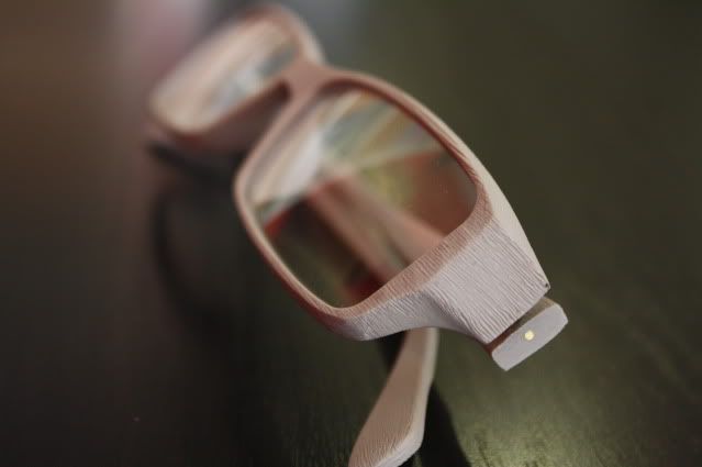
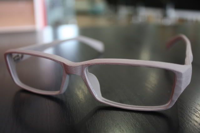
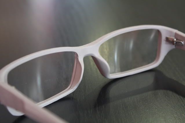
If your distance vision is still good, reading glasses can be an answer for presbyopia. They’re available in full-size lenses or in half-size glasses, as shown here.
Half-glasses have been designed to avoid a full lens – they allow the wearer to glance over the bottom reading portion for uncorrected distance viewing. The advantage of either half or full reading glasses (commonly referred to as ‘single vision’ glasses) is that they provide a large viewing area for up-close use. But the disadvantage is that they are limited to only one field of focus. Because they are only useful for near vision, they require you to keep taking them off when looking at a distance.
If you need correction for your near and far vision, you’ll need more than one pairs of glasses. Constant switching between these will be cumbersome and inconvenient.
OPTION 2 - ACCESS ENHANCED RANGE READING LENSES
Access lenses give you an ultra-wide reading area plus the wide, clear mid-range vision you can’t get from simple reading glasses. So everything looks clear from close-up to up to five feet away. Computer users and people doing similar work benefit from this type of correction.
The Access lens features a unique design that offers up to 50% more visual range than single vision reading lenses – so that everything from up close to arm’s length is seen clearly and comfortably.
Enhanced near vision for work or leisure.
Access lenses are ideal are ideal for almost any type of indoor activity at almost any distance.
Ask our staff about Access lenses. They can improve your view of the world in a blink.
OPTION 3 – BIFOCALS.
If you need visual correction for both distance and near vision, bifocals may serve both needs with the same lens. Invented as long ago as 1775 by Benjamin Franklin, bifocals now come in a variety of designs to fit various occupational needs and lifestyles. Their main advantage is that they eliminate the need to switch from on e pair of glasses to another.
In addition to being outdated technology, the disadvantages of bifocals are that the two fields of vision are separated by an obvious ‘line’. And even in bifocals where the line has been erased to look better (usually called ‘no-line,’ ‘blended,’ or ‘invisible’ bifocals), the change from near to far is abrupt and oftentimes annoying.
The biggest problem with bifocals of all types is that they do not solve the problems of intermediate vision – for instance, looking at a computer screen, reading prices on a supermarket shelf, or looking at a dashboard in the car. It can be very frustrating not to be able to see clearly at this often critical area between near and far.
OPTION 3 – TRIFOCALS.

Trifocals attempt to correct intermediate vision – that arm’s length distance such as your car dashboard or price labels on supermarket shelves. Trifocals include three distinct fields of vision in the same lens – one for distance, one for intermediate, and one for near. Each field is divided by a line. The chief advantage of trifocals is that they provide a third vision field.
But trifocals have two major disadvantages: the added line makes adjusting even more difficult than with bifocals, and they are not cosmetically appealing.
OPTION 4 - PROGRESSIVE LENSES.
Progressive lenses are a more recent development than bifocals and trifocals. They imitate the action of the human eye, providing the full range of focus from near to intermediate to fargradually, without lines, separations, or interruptions.
Progressives have no seams or lines, and look exactly like normal single vision lenses. Many people get used to them immediately; others need a slight period of adjustment. But once you are comfortable with them, it can be hard to imagine life without them.
Although they’re more expensive than other options, most wearers consider their eyes and vision to be well worth the extra investment. 92% of former bifocal wearers prefer Progressive Lenses and 97% successfully adapt to them.
Several companies currently manufacture progressives, examples being Essilor, Sola and Norville. At D`EYEWEAR Optical, we are not restricted to any particular supplier, using the best products available at very competitive prices.
Progressive lenses are available in a variety of plastic or glass materials to match lifestyle needs, as well as in a variety of tints and changeable (photochromic) tints.
The triple-patented Varilux Comfort design is based on a complex set of curves that vary from ellipses to parabolas to hyperbolas. It’s technology such as this that gives you better vision.

After having your eyes examined, you’ll be able to make an informed choice of eyeglass lenses in partnership with your eyecare professional will be glad to discuss how your needs can be met.
Remember, it’s advisable for everyone to have an eye examination at least once a year.
PRESCRIPTION SPECTACLES READY IN AROUND AN HOUR
At D`EYEWEAR Optical we believe in a quality, high speed service.
All our Practices are equipped with state of the art glazing laboratories and we hold a comprehensive range of single vision stock lenses including high index, aspheric and coated lenses.
This means many of our patients can have their new spectacles in around an hour.
If you have broken your spectacles we can also help.
We produce most single vision spectacles within an hour, repair broken spectacles and can even fit your own lenses into a new spectacle frame in an emergency.
How often have you visited you own Optician to be told that if you require lenses to your own frame you will have to be without your spectacles for a week or two?
At D`EYEWEAR Optical we use a high tech scanning system that allows us to order thin, cosmetically appealing lenses ready for your frames to be reglazed.
We advise you when your lenses are ready, arrange a convenient appointment for you to leave your spectacle frames and we will have them ready with new lenses in around an hour for you to collect.
What could be easier?


Single Vision lenses have only one viewing area throughout the lens. This corrected are
a can be for far distance, near distance or reading.
Bifocal Lenses have two distinct viewing areas within the same lens; the distance area and the near area. The distance area in bifocal is designed like a single vision lens, while the near area contains the distance prescription and the additional amount of ADD power nee
ded to see at a reading distance. If you would like to learn more about lined bifoca
ls


Trifocal lens is an extension of the bi-focals, which provide
vision correction only for distance and close up near viewing, leaving the int
ermediate area between distance and reading uncorrected. Tri-focals provide another segment on the lens above the reading viewing area to correct the intermediate viewing area between far and near.

Progressive lenses correct vision for two or three different distances without the
visible segment lines seen in bifocal or trifocal lenses. Instead they have a graduated section in which the power of the lens progresses smoothly from one prescription to the other, allowing the wearer to see clearly at all distances. Transition Zone is the area of progressive sunglasses lenses where the distance vision curve gradually changes into the near-vision curve.